We are pleased to report that a new article led by Caitlin Smith is now live and open access in Photoacoustics. Caitlin rigorously developed and validated a technique for photoacoustic vector-flow to measure the pixel-wise magnitude and direction of flowing optical absorbers. This work focused on ex vivo whole blood and a comparison to pulse-echo ultrasound. Caitlin’s work not only provides new capabilities for our lab, but for photoacoustic imaging as a whole. We look forward to the new directions and applications this work opens up! Well done, Caitlin!
Author: jjoh604
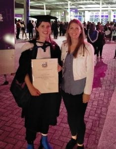
Congratulations to Caitlin on graduating with Honours and completing her thesis on photoacoustic velocimetry. After many months of waiting for an in-person graduation, Caitlin finally got to walk the plank this week! She has been such an asset to our biomedical imaging projects and we are thrilled she will be continuing her work in the PAL for her PhD.
A new publication hit the press this week on ultrasonic imaging of bone in Journal of Bone and Mineral Research PLUS. For the first time, we demonstrated quantitative measurements of blood flow in the cortical bone layer of a cohort of human volunteers. This paper is a continuation of Jami Shepherd‘s postdoctoral research in the Biomedical Imaging Laboratory in Paris, and ongoing collaborative work with colleagues in France and the Netherlands.
Mainstream photoacoustic imaging methods approximate the human body as a fluid. This assumption is reasonable for soft tissues, but breaks down for bones. Cortical bone is a stiffer material than soft tissues, with a wavespeed that is dependent on the direction of wave propagation (anisotropy). We have recently developed a new method for reconstructing photoacoustic images in collaboration with colleagues in the Laboratoire d’Imagerie Biomédicale in Paris, which accounts for the true properties of cortical bone. This work was published in Applied Physics Letters and serves as a launching point toward enabling photoacoustic imaging in human bones.