Photoacoustic imaging is a non-invasive modality that exploits the absorption-based contrast of optical imaging and high-resolution and deep wave propagation achievable with ultrasound. Potential clinical applications of this modality are widespread and include characterization of cancerous tumours and diseased blood vessels. In the Physical Acoustics Lab, we use an all-optical system to obtain broadband photoacoustic and laser-ultrasound images with a single setup. The photoacoustic image provides information about optical absorbers in tissue, such as lipids or hemoglobin; while laser-ultrasound is sensitive to acoustic scatterers like calcification.
Our research spans a wide range within photoacoustic imaging. First, we apply both known and novel methods of data acquisition and image reconstruction with roots in geophysics to improve the resolution and clarity of images. This is particularly relevant for imaging deep within inhomogeneous tissue, where scattering of acoustic waves can significantly limit quality. We are also developing non-contact acoustic detectors to expand potential clinical applications for photoacoustic imaging.
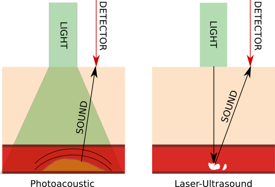
Photoacoustic generation and propagation (left) and laser-ultrasound generation and scattering (right).

Photoacoustic image (red) and laser-ultrasonic image (grey scale) of diseased artery. Layers of the artery wall (1-3) as well as calcification (4) are identified with arrows. A photoacoustic reflection artifact is denoted by a white arrow.
Related Publications:
- van der Neut, J., Johnson, J. L., van Wijk, K., Singh, S., Slob, E., Wapenaar, K. (2017). A Marchenko Equation for Acoustic Inverse Source Problems. The Journal of the Acoustical Society of America, 141, 4332-4346.
- Johnson, J. L., Shragge, J., van Wijk, K. Nonconfocal all-optical laser-ultrasound and photoacoustic imaging system for angle-dependent
deep tissue imaging. Journal of Biomedical Optics. 22 (4), 041014. - Johnson, J. L., van Wijk, K., Caron, J. N., and Timmerman, M. (2016). Gas-coupled laser acoustic detection as a non-contact line detector for photoacoustic and
ultrasound imaging. Journal of Optics, 18. - Johnson, J. L., van Wijk, K., Caron, James N., & Timmerman, M. (2016). Photoacoustic and ultrasound imaging with a gas-coupled laser acoustic line detector. Proc. of SPIE, paper 9708-38.
- Johnson, J. L, Shragge, J., van Wijk, K (2015). Image reconstruction of multi-channel photoacoustic and laser-ultrasound data using reverse time migration. Proc. of SPIE, Vol. 9223, paper 932314.
- Johnson, J. L., van Wijk, K., & Sabick, M. (2014). Characterizing Phantom Arteries with Multi-channel Laser Ultrasonics and Photo-acoustics. Ultrasound in Medicine & Biology, 40(3).
- Johnson, J., Sabick, M., & VanWijk, K. (2013). All-optical photoacoustic detection of absorbers in tissue phantoms. Journal of Medical Devices, 7(3).
- Johnson, J. L. (2013). Toward Characterization of Diseased Vascular Structures Using Noncontact Photoacoustic and Laser-Ultrasound Imaging: A Phantom Study. (Master’s thesis, Boise State University).