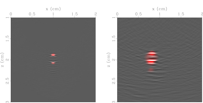
The latest issue of the Journal of Biomedical Optics features an article led by Jami Johnson, in collaboration with our good friend Jeffrey Shragge (from the University of Western Australia). In it, we apply some the latest tricks stemming from seismology to medical imaging of arteries. This particular publication focuses on theory and numerical simulations, but below you can see the first scan of an ex-vivo artery in the Physical Acoustics Lab!
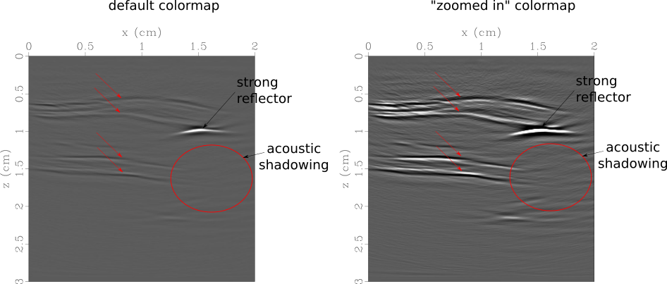
Ultrasound along the length of an ex-vivo carotid artery. The top and bottom walls of the vessel are clearly defined by two reflectors each. On the right, the top vessel wall increases in thickness and calcification is seen on the bottom of the top wall.
Comments are closed.